By Elaine J Wabwire
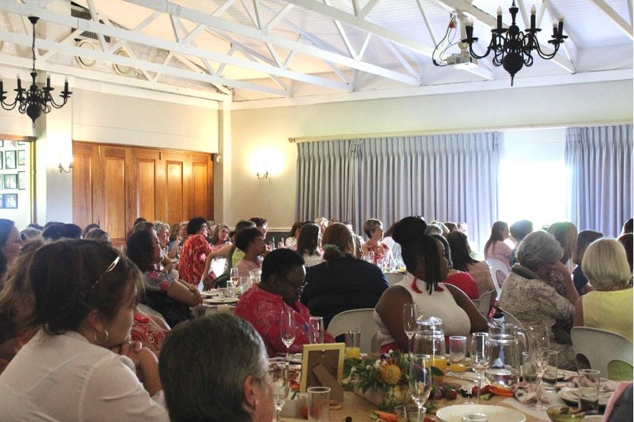
The Makhanda Hospice hosted its 14th annual breast cancer lunch on 20 October 2023 at the Wyvern Club in Kingswood College. The event focused on generating support for and raising critical funds for hospice and spreading awareness about the importance of breast cancer research and support.
The event started with an opening address by Janine Peinke, Practice manager at Makhanda and Sunshine Coast Hospice, shedding light on the work done by the Makhanda Hospice. The hospice has a staff of seven registered nurses, nine carers and one social auxiliary worker offering holistic care that encompasses emotional, social, spiritual and medical support to patients in Makhanda, Bathurst, Kenton-on-Sea, Port Alfred and Alexandria, supporting approximately 130 patients monthly. “By supporting this event today, you are helping us to keep providing that vital service to those who need it,” Peinke said.

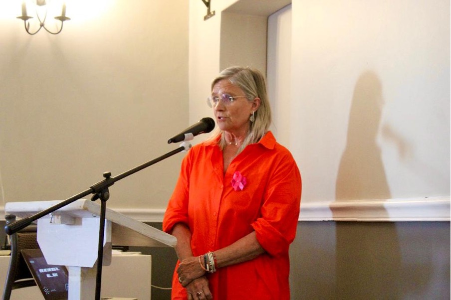
Sandy Coffey, a guest speaker at the event and a Life Coach and photographer, delivered an inspirational address. She spoke about the reality of imagination and encouraged the ladies to embark on a journey of discovery and empowerment through creativity. Coffey introduced the audience to the concept of “funterventions”, a practice aimed at infusing daily life with moments of unaltered joy. In pursuing creativity, she encourages the audience to “fly against convention and to be fearless.”
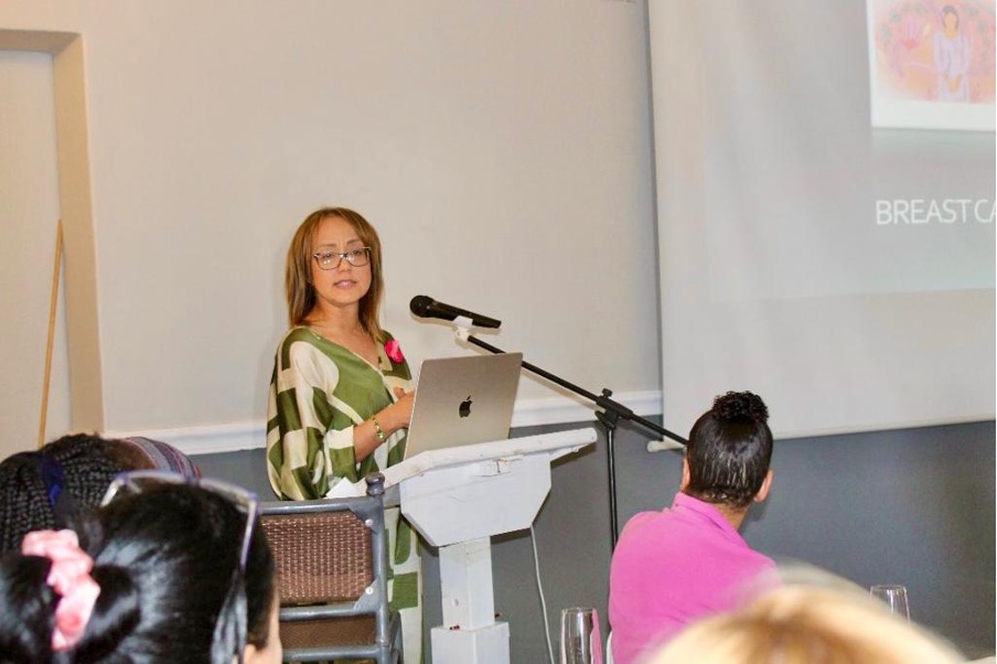
In light of breast cancer awareness month, Dr Sharon de Kock, a Radiologist from Bay Radiologist, spoke about the importance of early detection and regular screening for early detection of breast cancer. She outlined the risk factors associated with breast cancer and the significance of identifying individuals who should undergo screening. She outlined the risk factors that play a role in breast cancer:
- Gender: Being female, breast cancer is a hormonal disease; the hormone oestrogen plays a significant role in breast cancer development.
- Genetic factors: Play a pivotal role in our susceptibility to breast cancer. The presence of breast cancer genes, including the BRCA genes, acts as a safeguard against cancer. However, when these genes mutate, our risk of cancer increases. Specifically, mutations in the BRCA 1 AND 2 genes.
- Radiation exposure: Previous exposure to the chest wall may increase your chances of getting breast cancer.
- Family history: When close relatives, particularly on the maternal side, have experienced breast cancer at a young age, the likelihood of inherited susceptibility increases. Dr de Kock added that “Cancers seen at an advanced age are not necessarily related to family history, but the earlier the cancer, 30-40, you tend to be screened earlier.”
- Breast density: Breast density is an essential factor in breast cancer screening. Breasts are made up of a mixture of glandular tissue and fat. Breasts with a higher proportion of glandular tissue tend to appear denser on mammograms, making it harder to detect early stages of breast cancer.
- Early Menstruation Starting your menstrual cycle at a young age, which leads to prolonged exposure to oestrogen, can increase the risk of breast cancer.
- Lifestyle-related factors like a diet high in saturated fats and low in fruits and vegetables and physical inactivity
Dr de Kock emphasized the need for tailored screening based on individual risk factors. High-risk individuals are patients whose mothers, sisters, and daughters had breast cancer, especially at an early age. These individuals are screened earlier.
Recognising the unique needs of these individuals, Dr de Kock stressed the importance of a modified screening approach. In this case, Mammograms involving radiation exposure are often replaced by ultrasounds to prevent radiation exposure. Dr de Kock said, “We don’t necessarily do mammograms on them. We start with ultrasounds because they do not use radiation exposure. We would do yearly check-ups.” She added, “High-risk patients require regular follow-ups but not always with mammography.” A combination of ultrasounds and, in the case of very dense breasts, MRI scans are recommended every second or fifth year. This approach allows radiologists to follow up very closely and observe any changes or abnormalities in the breast.
Average-risk individuals should start annual mammography from the age of 30, with the frequency depending on the density of the breast. From then on, every second year is recommended for a person whose mammogram was normal if the results were normal. We do a normal screening from age 35 onwards; if there is no clinical indication for you to come earlier, then we can see you at 40 years.
Ultrasound helps create an image while a mammogram gives us a more precise image to see inside the breast and what is going on. “The denser your breasts are, the easier it is to mask what is happening in your breast.” Ultrasounds use crystals to create a picture; there is no radiation involved in an ultrasound. Dr de Kock highlighted that the major issue with ultrasounds for screening is that “We cannot see the calcifications, and these are the precursors of breast cancer. An ultrasound will show us if there is a lump in your breast, but we cannot see the precursors, the microcalcifications of cancer with an ultrasound”.